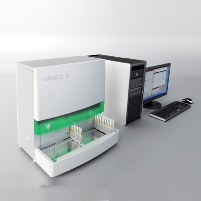
Urised 3
Documentation / Downloads

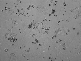
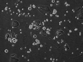
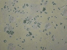
UriSed 3 is a professional automated urine sediment analyzer with a revolutionary new optical system combining bright-field and phase contrast microscopy. It offers a uniquely advanced method of visualization and recognition of formed elements in urine samples. By automating the traditional gold standard method of sediment analysis it increases reliability, improves work-flow and decreases turnaround time.
This instrument belongs to the Facelift generation of the UriSed technology. Its operation is based on the same patented measurement technique, which is actually the automation of traditional manual microscopy. Working without any special liquid reagents UriSed 3 performs sample preparation, takes multiple images of each sample through a built-in microscope, and evaluates the images using the Auto Image Evaluation Module (AIEM), a high-quality image processing software.
The UriSed 3 microscopic urine sediment analyzer is a stand-alone instrument, which can be connected to the LabUMat 2 urine analyzer. Together, the two instruments make up a
Complete Urine Laboratory System.